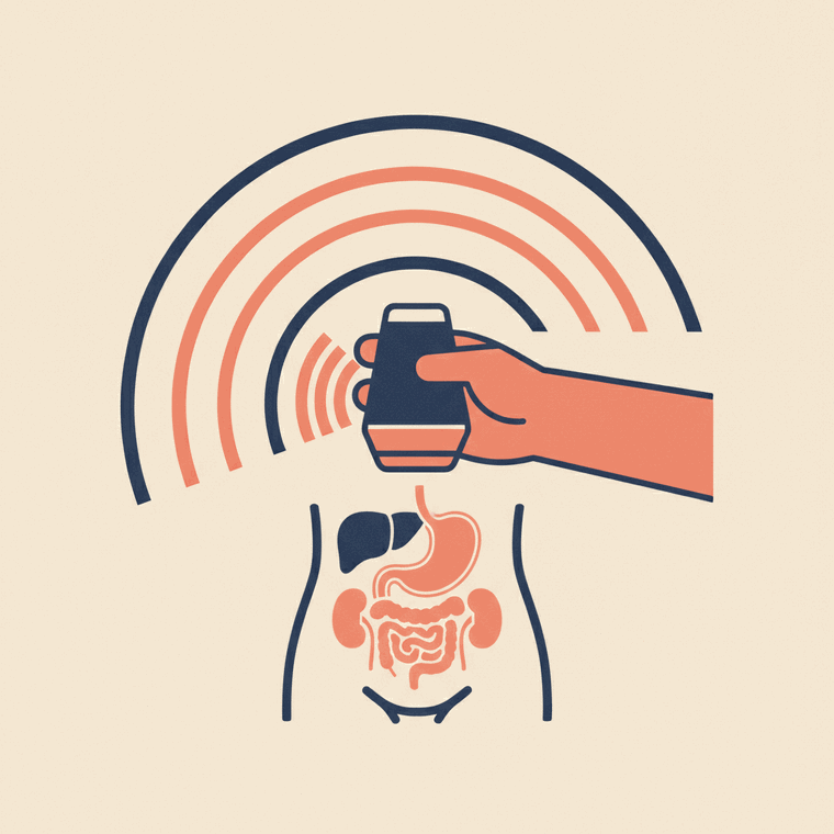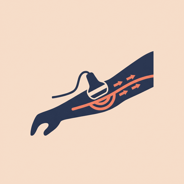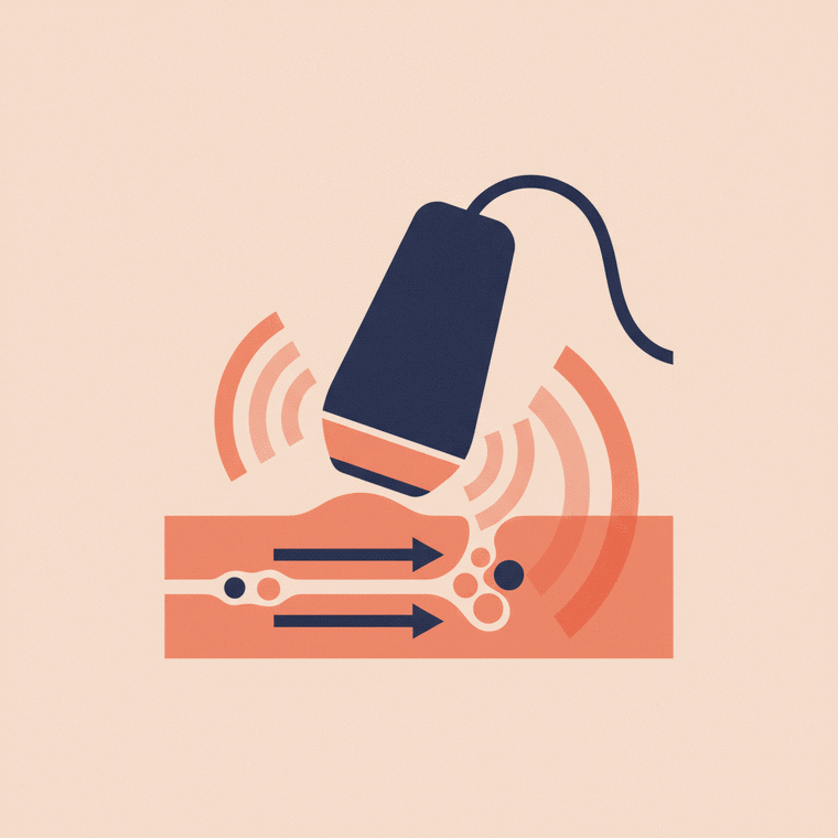
A Doppler ultrasound measures the direction and speed of blood moving through your vessels — named after the physicist Christian Doppler, whose effect it relies on. It's the same machine as an ordinary ultrasound, with the blood-flow function switched on, so it uses no radiation. In Australia it's most often used to look for a clot in a leg vein (DVT), to check narrowed arteries in the legs or neck, or to check blood flow to a baby in pregnancy.
Key takeaways
- Doppler is ordinary ultrasound with the blood-flow function on — no radiation, no injection.
- It's also called duplex, vascular or colour-flow ultrasound. They mean the same thing.
- Which scan you have depends on the question: leg veins, leg or neck arteries, abdomen, or pregnancy.
- A positive leg-clot result is acted on the same day.
- Reports use set phrases — 'fully compressible', 'triphasic', 'no significant stenosis' — that have specific meanings.
You’ve probably seen the flashes of red and blue on an ultrasound screen, or heard a rhythmic whooshing during a scan. That’s Doppler — the part of ultrasound that measures blood flow. If you’ve been booked for one, this page covers which scan you’re likely having, what the report will say, and what happens after.
What a Doppler ultrasound is — and what “Doppler” means
Ordinary ultrasound shows structure: what a vessel looks like. Doppler adds movement — the direction and speed of blood as it flows.[1] It works on the Doppler effect, named after the Austrian physicist Christian Doppler: the pitch of a sound wave changes when it bounces off something moving, and a computer turns that shift into a picture of flow.
It’s the same probe and the same machine as any other ultrasound, with the flow function switched on. That means no radiation and no injection.
You may see it called duplex ultrasound, vascular ultrasound, colour-flow ultrasound or just “a Doppler”. For practical purposes these all describe the same thing.
Which Doppler scan have you been booked for?
“Doppler ultrasound” isn’t one test. The vessel being looked at depends on the question your doctor is asking, and that changes what happens on the day.
| Scan | What it checks | Usually ordered because |
|---|---|---|
| Venous Doppler (legs) | Leg veins, for a clot | Swollen, painful or red calf; after surgery or a long flight |
| Arterial Doppler (legs) | Leg arteries, for narrowing | Calf or thigh pain when walking that eases with rest; a slow-healing foot wound; diabetes |
| Carotid Doppler (neck) | Neck arteries to the brain | After a TIA or stroke; a sound heard in the neck with a stethoscope |
| Renal or abdominal Doppler | Blood flow to kidneys, liver or bowel | High blood pressure that’s hard to control; abnormal liver tests. Fasting is usually needed |
| Obstetric Doppler | Blood flow in the placenta and baby | A baby measuring small; high blood pressure in pregnancy; going past your due date |
| Scrotal Doppler | Blood flow to the testicle | Sudden severe pain — this is treated as urgent, same day |
If you’re not sure which you’re having, the referral will name the body part. The one that most often needs preparation is the abdominal or renal scan.
What happens during the scan
Warm gel goes on the skin and the sonographer moves the probe over the area. For a leg vein scan they will press firmly at intervals — this is the point of the test, not carelessness: a healthy vein squashes flat under gentle pressure and a vein with a clot in it doesn’t.[3]
For arterial studies you may also have blood-pressure cuffs placed on the thigh, calf and ankle to compare pressures down the limb.
Expect 20 to 45 minutes, longer than a standard ultrasound because each vessel is checked in turn. You’ll hear a loud whooshing timed with your pulse — that’s spectral Doppler turning flow into sound. Most scans need no preparation and no aftercare, and you can drive and go back to work straight afterwards. Abdominal and renal Doppler are the exception: you’ll usually be asked not to eat for several hours so bowel gas doesn’t obscure the vessels.
Is it safe?
Yes. Doppler uses sound waves, not ionising radiation, so there’s no dose to account for and no radiation risk to weigh up.[2] It’s used routinely in pregnancy for that reason. Ultrasound does deposit a very small amount of energy as heat, so operators follow output limits and keep scanning time to what’s clinically needed — a background safeguard rather than something you need to think about. There is no recovery period.
Reading your Doppler report: the words you’ll see
Doppler reports are written for your referring doctor, in a fairly fixed vocabulary. Knowing the phrases makes the report far less alarming to read.
Phrases that indicate a normal study
- “Fully compressible” — the vein flattens completely under the probe. This is the key finding that argues against a clot.
- “No evidence of deep vein thrombosis” — no clot seen in the veins examined.
- “Triphasic” or “biphasic” waveform — the normal shape of arterial flow in a limb. Triphasic is the healthiest pattern.
- “Patent” — the vessel is open and flowing.
- “No haemodynamically significant stenosis” — any narrowing present isn’t tight enough to meaningfully reduce flow.
- “Antegrade flow” — blood moving in the expected direction.
Phrases that indicate something was found
- “Occlusive” vs “non-occlusive” thrombus — whether the clot blocks the vein completely or partly.
- “Acute” vs “chronic” — a fresh clot versus old, scarred vein from a previous one. These are managed very differently.
- “Monophasic” or “dampened” flow — arterial flow has lost its normal shape, suggesting narrowing further upstream.
- “Reflux” — blood flowing backwards through a valve that isn’t closing. Common, and usually about varicose veins rather than anything dangerous.
- “Peak systolic velocity” (PSV) — the fastest speed measured at a point. Blood speeds up through a narrowing, so a high PSV suggests a tighter narrowing.
- “Stenosis” with a percentage range — an estimate of how narrowed an artery is, for example 50–69% or 70–99%.
One important caveat about the numbers. The velocity thresholds used to convert a speed into a percentage narrowing are not universal — different laboratories and guidelines use different criteria, and a good report will state which criteria were applied. That’s why a figure from your report can’t be compared directly against one from another clinic, and why the percentage band matters more than the raw number.
A Doppler is also always read alongside your symptoms. A radiologist interprets the images and traces the sonographer captured, and reports to the doctor who referred you.
What happens next, by result
- Normal scan, symptoms settling — usually nothing further. If your symptoms persist or worsen, go back to your doctor; a scan is a snapshot of one moment.
- Clot found in a leg vein — this is acted on the same day. Your referring doctor or the emergency department is contacted directly, and treatment with blood-thinning medication is usually started promptly.
- Below-knee or inconclusive result — clots in the smaller calf veins can be genuinely hard to see. A repeat scan in about a week is a standard and appropriate next step, not a sign anything was done badly.
- Significant arterial narrowing — referral onward, typically to a vascular surgeon, or to a neurologist or stroke service for the neck arteries.
If your report says a clot was found and you can’t reach your referring doctor, don’t wait for the next available appointment — go to an emergency department. Sudden breathlessness or chest pain after a leg clot needs immediate emergency care.
Cost, Medicare and bulk billing
Like other imaging in Australia, a Doppler ultrasound attracts a Medicare rebate only if you have a valid referral from a GP or specialist. Without one there’s no rebate, and you pay the full fee.
Whether you pay anything on the day depends on the practice, not on the scan. Providers set their own fees: some bulk bill vascular ultrasound outright, others charge a gap. Scans done in a public hospital, or as an inpatient, are generally covered.
It’s worth ringing ahead and asking three things: whether they bulk bill this specific scan, what the out-of-pocket cost will be if not, and whether that changes with a concession or pension card. Our guide to what a scan really costs in Australia explains how the rebate, the gap and the Medicare Safety Net fit together.
How long results take, and getting a copy
For a routine outpatient Doppler, the report usually reaches your referring doctor within a day or two. Urgent scans — a suspected DVT, a painful testicle — are read and phoned through immediately. Scans done in an emergency department are typically reported the same visit.
You’re entitled to a copy of your own images and report; ask the practice at the time of the scan. More on this in getting your ultrasound results.
Doppler in pregnancy
Obstetric Doppler checks blood flow through the placenta and umbilical cord, usually when there’s a question about the baby’s growth or your blood pressure. It’s a different scan with its own findings and thresholds — see Doppler ultrasound in pregnancy.
Frequently asked questions
Why does the sonographer keep pressing on my leg?
That pressing is the test. A normal vein flattens completely under gentle pressure from the probe; a vein containing a clot stays open. It shouldn’t be more than briefly uncomfortable — say so if it is.[3]
Can a Doppler miss a clot?
It can, particularly in the smaller veins below the knee, which is why a repeat scan after about a week is standard when the first is inconclusive but symptoms continue. If your leg gets worse, or you become breathless, seek care again rather than relying on the earlier scan.[3]
Do I need to fast?
Only for abdominal or renal Doppler, where bowel gas can obscure the vessels. Leg, neck and pregnancy scans need no preparation.
What do the red and blue colours mean?
They show the direction of flow relative to the probe, and the shade reflects speed — not “arteries versus veins”, and not “good versus bad”. A vessel can change colour on screen simply because it curves.[1]
What’s the difference between a Doppler, a duplex and a vascular ultrasound?
In everyday use, nothing. “Duplex” strictly means combining the grey-scale picture with flow measurement, which is what almost every Doppler scan does.
Can I drive and work afterwards?
Yes. There’s no sedation, no injection and no aftercare, so you can go straight back to normal activity.
Sources
- RadiologyInfo.org (RSNA & ACR) — General Ultrasound (Doppler) — www.radiologyinfo.org/en/info/genus
- RANZCR / InsideRadiology — Ultrasound — www.insideradiology.com.au/ultrasound-hp/
- healthdirect Australia — Deep vein thrombosis (DVT) — www.healthdirect.gov.au/deep-vein-thrombosis