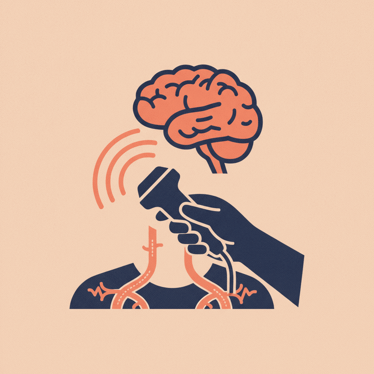
A carotid Doppler is a vascular ultrasound of the carotid arteries in your neck — the main vessels supplying blood to the brain. It checks for narrowing (usually from fatty plaque build-up) that can raise the risk of stroke. It uses sound waves, not radiation, is painless, and needs no preparation. It's often requested after a mini-stroke (TIA) or to assess stroke risk.
Key takeaways
- A carotid Doppler checks the neck arteries that supply the brain.
- It looks for narrowing from plaque that can raise stroke risk.
- It uses sound waves and shows blood flow — no radiation.
- It's painless and needs no preparation.
A carotid Doppler focuses on the carotid arteries — the two main blood vessels running up either side of your neck to supply the brain.[1] It uses the Doppler technique to show blood flow, with no radiation.
What it’s for
Over time, fatty deposits (plaque) can build up and narrow the carotid arteries, which can raise the risk of stroke.[1] A carotid Doppler checks for this narrowing and measures how much it’s affecting blood flow. It’s commonly requested:
- After a mini-stroke (TIA) or stroke symptoms
- To assess stroke risk in people with risk factors
- To follow up a narrowing found earlier, or before certain surgery
By showing the speed and turbulence of flow, it can estimate how significant a narrowing is — which guides whether medication or a procedure is needed.[1]
What to expect
You lie back with your neck slightly extended while a sonographer applies gel and moves the probe over each side of your neck.[1] You may hear the pulsing blood flow. It’s painless, needs no preparation, and has no aftercare. A full study takes around 20–30 minutes.
Frequently asked questions
Why would I need a carotid Doppler?
To check whether the neck arteries supplying your brain are narrowed — important for assessing and managing stroke risk, especially after warning symptoms like a TIA.[1]
Is it uncomfortable?
No — it’s painless. A probe is moved gently over your neck with gel; you may hear your blood flow.[1]
What happens if it finds a narrowing?
Your doctor interprets the result with your overall risk. Depending on how significant the narrowing is, that might mean medication and risk-factor management, or in some cases a procedure to reduce stroke risk.[1]
Sources
- RadiologyInfo.org (RSNA & ACR) — General Ultrasound (Doppler) — www.radiologyinfo.org/en/info/genus
- RANZCR / InsideRadiology — Ultrasound (Coombs) — www.insideradiology.com.au/ultrasound-hp/