An ultrasound (sonography) scan uses high-frequency sound waves — not radiation — to create live, moving pictures of the inside of the body. A hand-held probe sends sound in and listens for the echoes, which a computer turns into an image in real time. It's painless, radiation-free and extremely safe — the go-to scan in pregnancy and a first-line test for the abdomen, thyroid, blood vessels and more. Its main limit is that sound can't pass through bone or gas.
Key takeaways
- Ultrasound uses sound waves and shows images in real time — no radiation.
- It's the safest imaging test, and the standard for monitoring pregnancy.
- A probe and gel are moved over the skin; it's painless and needs no aftercare.
- Sound can't pass through bone or gas, which limits what it can see.
Ultrasound is the friendliest scan in the department: no radiation, no tunnel, no injection — just a probe, some gel, and a live picture on a screen. Here’s how it works and what it’s used for.
How ultrasound works
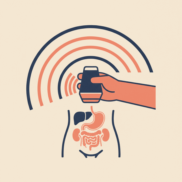
Ultrasound uses the same principle as the sonar used by ships and bats. A hand-held probe (a transducer) sends inaudible, high-frequency sound waves into the body. When the waves hit organs, fluid and tissue, they bounce back as echoes, and the probe listens for them.[1] A computer measures the echoes — how strong they are and how long they take to return — and instantly builds a real-time picture on the monitor.[1]
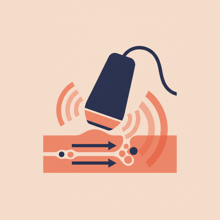
Because it works with sound rather than radiation, ultrasound is extremely safe, and the live image lets the operator watch things move — a beating heart, a moving baby, or blood flowing through a vessel.
What ultrasound is used for
It’s a genuine all-rounder, used to investigate pain, swelling and infection and to examine many organs, including:[1]
- The abdomen — liver, gallbladder, kidneys, pancreas, spleen and the aorta
- The pelvis — bladder, uterus and ovaries
- Pregnancy — monitoring the baby’s growth and wellbeing
- The thyroid and neck, breast lumps, and the testes
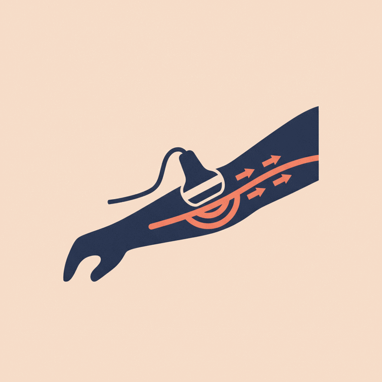
- Blood vessels — checking blood flow with the Doppler technique, including leg veins for clots
- The heart — an “echocardiogram” (echo)
- Guiding procedures — steering a needle in real time for a biopsy or drainage
In babies, it can even see the brain, hips and spine, because their bones are softer.[1]
What a scan is like
A sonographer applies gel to the skin and moves the probe over the area, watching the image live.[1] It’s painless (though pressure over a tender area may be briefly uncomfortable), takes about 30 minutes, and the gel wipes off easily afterwards.[1] You can return to normal activities straight away.
Some scans use an internal probe for a closer view — a transvaginal ultrasound for the uterus and ovaries, or a transrectal one for the prostate.[1] Your clinic will explain if that applies.
Its limits
No scan does everything. Sound waves are disrupted by air or gas and can’t pass through bone, so ultrasound isn’t ideal for the gas-filled bowel, the air-filled lungs, or seeing inside bones.[1] It’s also harder in larger patients, where sound has further to travel. In those cases, CT or MRI may be used instead.
Frequently asked questions
Is an ultrasound safe?
Yes — standard diagnostic ultrasound has no known harmful effects and uses no radiation, which is exactly why it’s the preferred scan in pregnancy.[1]
Does an ultrasound hurt?
No — it’s painless. You’ll feel the probe and cool gel moving over your skin; pressure over a sore area might be briefly uncomfortable.[1]
Why does the sonographer press so hard sometimes?
A little pressure helps the sound waves reach deeper structures and can move gas out of the way. It shouldn’t be painful — tell the sonographer if it is.[1]
Sources
- RadiologyInfo.org (RSNA & ACR) — General Ultrasound — www.radiologyinfo.org/en/info/genus
- RANZCR / InsideRadiology — Ultrasound (Coombs) — www.insideradiology.com.au/ultrasound-hp/
- healthdirect — Ultrasound — www.healthdirect.gov.au/ultrasound