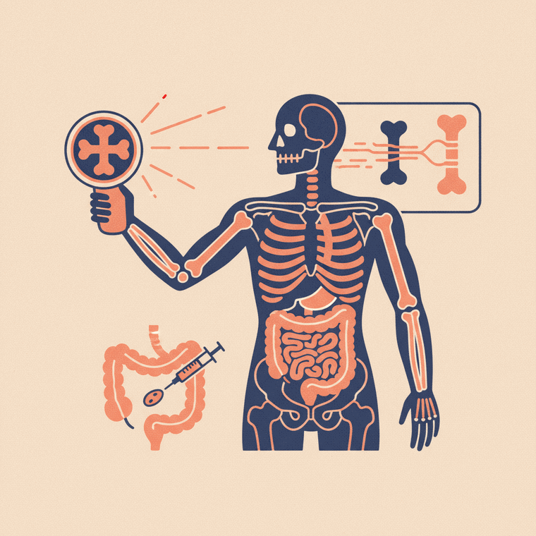
Fluoroscopy (real-time X-ray) is used two ways. For diagnosis, it watches the body in action — barium studies of the swallowing tube, stomach and bowel. For guiding procedures, it steers instruments precisely in real time — angiograms (imaging blood vessels with contrast), joint injections and arthrograms, and placing or checking catheters, drains and lines. Most of these use a contrast agent, and the radiation dose varies with how long the procedure takes.
Key takeaways
- Diagnostic uses: barium studies of the gut (swallowing, stomach, bowel).
- Procedure guidance: angiograms, joint injections, catheter and line placement.
- Most fluoroscopy uses a contrast agent (barium or iodine).
- Radiation dose varies — longer procedures use more.
Fluoroscopy’s live, moving X-ray view makes it useful across a surprising range of situations — both to diagnose problems and to guide treatment. Here’s the full picture.
Diagnostic uses: watching the body work
By showing movement in real time, fluoroscopy can assess things a still image can’t:[1]
- Barium swallow — watching the throat and oesophagus as you swallow, for swallowing difficulty or reflux.
- Barium meal and follow-through — studies of the stomach and small bowel.
- Barium enema — imaging the large bowel (less common now that colonoscopy is widely used).
These use a barium or iodine contrast so the digestive tract shows up clearly.[2]
Procedure guidance: steering with precision
This is where fluoroscopy does much of its modern work — letting a doctor watch an instrument move inside the body and place it exactly:[1]
- Angiograms — imaging blood vessels by injecting iodine contrast through a catheter and watching it flow, to find narrowing, blockages or aneurysms.
- Joint injections and arthrograms — guiding a needle precisely into a joint for an injection or to inject contrast before an MRI or CT.
- Placing and checking catheters, drains and lines — for example a central line or a feeding tube.
- Some pain-management injections into the spine.
Contrast and radiation
Most fluoroscopy procedures use a contrast medium, so the usual contrast precautions apply (tell staff about allergies and kidney problems).[2] Because the beam is on for longer, a long, complex procedure can approach the dose of an extensive CT, while a short study is a modest dose.[3] The team uses only the radiation needed, and it’s generally avoided in pregnancy unless necessary.
Frequently asked questions
Is fluoroscopy only for the digestive system?
No — barium studies of the gut are one use, but a lot of fluoroscopy is about guiding procedures, such as angiograms, joint injections and placing catheters, anywhere in the body.[1]
What’s an angiogram?
An angiogram is a fluoroscopy study of the blood vessels: iodine contrast is injected and watched in real time as it flows, to find narrowing, blockages or aneurysms.[1]
Does fluoroscopy hurt?
The imaging itself is painless. If it’s guiding a procedure — an injection or catheter — you may feel that part, and local anaesthetic is often used. Your team will explain what to expect.[1]
Sources
- RadiologyInfo.org (RSNA & ACR) — Fluoroscopy — www.radiologyinfo.org/en/info/fluoroscopy
- RANZCR / InsideRadiology — Iodine-containing contrast medium (ICCM) — www.insideradiology.com.au/iodine-containing-contrast-medium/
- RANZCR / InsideRadiology — Radiation risk of medical imaging — www.insideradiology.com.au/radiation-risk/