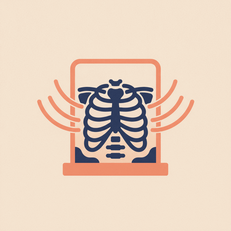
A chest X-ray is a fast, very low-dose scan of the heart and lungs. It's used to investigate a cough, breathlessness, chest pain or infection, to look for pneumonia or fluid, and to check the size of the heart. The dose is tiny — about 0.02 mSv, roughly two to three days of natural background radiation — and there's no preparation and no aftercare.
Key takeaways
- A chest X-ray quickly images the heart and lungs.
- Used for cough, breathlessness, chest pain, infection and heart size.
- Very low dose — about 0.02 mSv (a few days of background radiation).
- No preparation, no aftercare; you may be asked to hold a breath.
The chest X-ray is one of the most common scans there is — a quick, simple look at the heart and lungs that answers a surprising number of questions.
What a chest X-ray shows
It’s used to assess the heart and lungs and the space around them.[1] Common reasons include:[1]
- A cough, breathlessness or chest pain
- Suspected pneumonia or other lung infection
- Fluid on or around the lungs
- Checking the size and outline of the heart
- Following up a known condition, or before some operations
Because bone, air and soft tissue in the chest absorb the beam so differently, they stand out clearly from one another — making the chest one of the areas X-ray does best.
How safe is it?
Very. A chest X-ray delivers only about 0.02 mSv of radiation — roughly two to three days of the natural background radiation we all receive anyway, with no short-term effects.[3] The benefit of the information far outweighs this tiny risk. If you are, or might be, pregnant, tell the team — though a chest X-ray is away from the abdomen and very low dose.[2]
What to expect
There’s no preparation. You’ll usually stand with your chest against a flat plate, and the radiographer may ask you to take a deep breath and hold it so your lungs are fully inflated for a clear picture.[2] It takes only a moment, there’s no aftercare, and you can carry on straight away.
Frequently asked questions
Is a chest X-ray dangerous?
No — the dose is tiny (about a few days of natural background radiation), with no short-term effects. The benefit almost always outweighs the very small risk.[3]
Why am I asked to hold my breath?
Holding a deep breath fully inflates the lungs, which gives a clearer, more useful picture of the lungs and heart.[1]
Can a chest X-ray detect lung cancer?
It can show some lung abnormalities, but it’s not a reliable screening test for lung cancer — small or early cancers can be missed. If lung cancer is a concern, your doctor may arrange a CT instead.[1]
Sources
- RadiologyInfo.org (RSNA & ACR) — Chest X-ray — www.radiologyinfo.org/en/info/chestrad
- RANZCR / InsideRadiology — Plain radiograph / X-ray — www.insideradiology.com.au/plain-radiograph-x-ray-hp/
- ARPANSA — Medical imaging radiation doses — www.arpansa.gov.au/understanding-radiation/radiation-sources/more-radiation-sources/medical-imaging
- healthdirect — X-rays — www.healthdirect.gov.au/x-rays