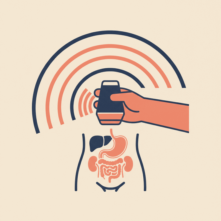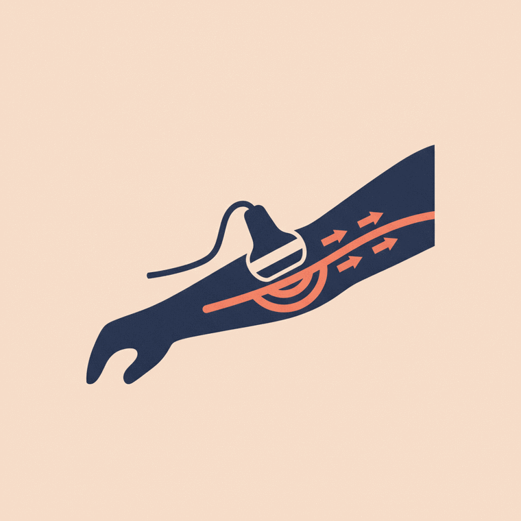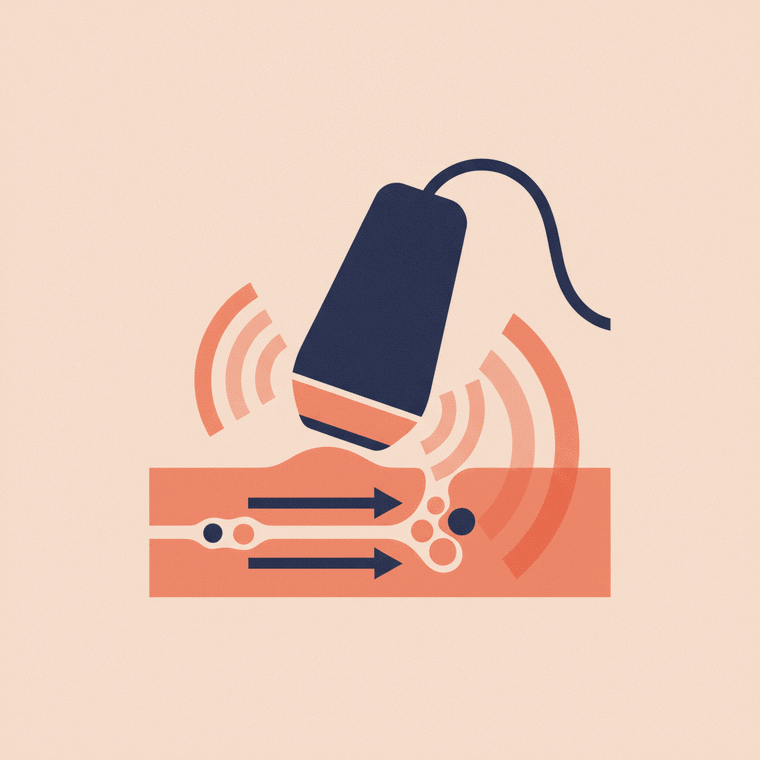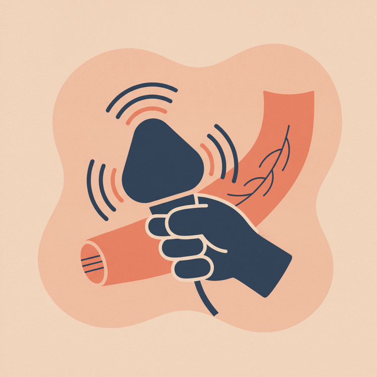
Vascular ultrasound uses sound waves — and the Doppler technique — to image your blood vessels and measure blood flow, with no radiation. It's used to look for blood clots in veins, narrowed or blocked arteries, varicose veins, and the blood supply to organs. Doppler shows the direction and speed of flow in real time, so it can reveal problems a still picture would miss. It's painless and needs no aftercare.
Key takeaways
- It images arteries and veins and measures blood flow using Doppler — no radiation.
- Used for clots (DVT), narrowed arteries, varicose veins and organ blood supply.
- Doppler shows the direction and speed of blood flow in real time.
- It's painless with no aftercare.
Vascular ultrasound is the use of ultrasound to examine your blood vessels — both arteries and veins. It combines a normal ultrasound image with the Doppler technique, which measures the direction and speed of blood flow in real time, all with no radiation.[1]
How it works
A hand-held probe sends sound waves into the body. For the flow measurements, it uses the Doppler effect — the change in pitch of the sound waves as they bounce off moving blood cells — which a computer turns into colours or graphs showing how blood is moving.[1] You may even hear a rhythmic “whooshing” as the flow is detected.
What it’s used for
Vascular ultrasound can help find:[1]
- Blood clots in the deep veins of the legs (deep vein thrombosis, or DVT)
- Narrowed or blocked arteries affecting circulation
- Varicose veins and how the leg veins are working
- The blood supply to organs, grafts or a transplant
- Reduced, absent or increased blood flow — which can point to a blockage, or to infection
Because it shows flow as it happens, it can reveal problems that a still picture would miss — and it helps doctors decide, for example, whether someone is a good candidate for a procedure to open a narrowed artery.[1]
What to expect
A sonographer applies gel and moves the probe over the area; it’s painless, takes around 30–45 minutes for a full study, and needs no aftercare.[1] For leg or neck vessels, you’ll simply lie back while each side is checked.
Its limits
As with all ultrasound, sound doesn’t travel well through bone or gas, and very deep vessels can be harder to assess in larger patients — in which case CT or MRI angiography may be used instead.[1]
Frequently asked questions
Does a vascular ultrasound hurt?
No — it’s painless. The sonographer moves a probe over the skin with gel; you may hear the whooshing sound of your blood flow.[1]
How does it show blood flow?
It uses the Doppler technique, which measures how the sound waves change as they bounce off moving blood — giving the direction and speed of flow.[1]
Sources
- RadiologyInfo.org (RSNA & ACR) — General Ultrasound (Doppler) — www.radiologyinfo.org/en/info/genus
- RANZCR / InsideRadiology — Ultrasound (Coombs) — www.insideradiology.com.au/ultrasound-hp/