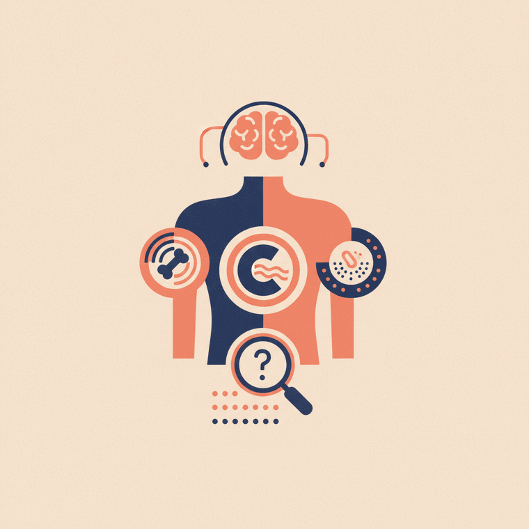
Common examples: a suspected broken bone → X-ray; pregnancy → ultrasound; gallstones → ultrasound; a suspected stroke → CT then often MRI; a knee or spine problem → MRI; cancer staging → PET-CT; osteoporosis → DXA. Each matches the scan to what needs to be seen, while keeping radiation as low as possible.
Key takeaways
- Broken bone → X-ray; pregnancy and gallstones → ultrasound.
- Suspected stroke → CT, often followed by MRI.
- Knee, spine and brain soft-tissue problems → MRI.
- Cancer staging → PET-CT; osteoporosis → DXA.
Imaging makes much more sense with real examples. Here are common situations and the scan usually chosen — and why.
Everyday examples
- A suspected broken wrist → X-ray. Quick and excellent for bone; the first choice for most fractures.[1]
- Pregnancy → ultrasound. Sound waves, no radiation — the safe mainstay for monitoring a baby.
- Suspected gallstones or abdominal pain → ultrasound first, sometimes a CT for more detail.
- A suspected stroke → CT quickly (to rule out a bleed), often followed by MRI for finer detail of the brain.
- A knee, shoulder or spine problem → MRI. Superb for soft tissues like cartilage, ligaments and discs.
- Cancer staging → PET-CT, showing how active a cancer is and whether it has spread.
- Osteoporosis check → DXA, a low-dose scan measuring bone density.
The pattern behind the choices
Notice the logic:[2]
- Bone problems lean on X-ray and CT.
- Soft-tissue problems lean on MRI and ultrasound.
- Function questions (how active or how well something works) lean on nuclear medicine / PET.
- Where possible, the radiation-free option (ultrasound or MRI) is preferred — especially for children and in pregnancy.
Why the first scan isn’t always the last
Often one scan leads to another: an ultrasound might prompt an MRI, or a CT might be followed by a targeted study. This step-by-step approach uses each scan’s strengths and avoids unnecessary radiation.[2]
Frequently asked questions
Why an X-ray for a broken bone but an MRI for a knee ligament?
X-ray is excellent for bone but shows soft tissue poorly. A torn ligament is soft tissue, which MRI shows in detail — so each problem is matched to the scan that sees it best.[1]
If I have belly pain, which scan will I get?
Often an ultrasound first (no radiation, good for gallstones and many organs), with a CT if more detail is needed — for example for appendicitis or in an emergency. Your doctor decides based on your symptoms.[1]
Sources
- RadiologyInfo.org (RSNA & ACR) — Tests & Treatments — www.radiologyinfo.org/en/test-treatment
- RANZCR / InsideRadiology — Radiation risk of medical imaging — www.insideradiology.com.au/radiation-risk/