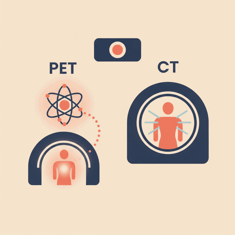
A CT shows detailed structure (what the body looks like) using X-rays. A PET shows function (how active tissues are) using a radioactive tracer. They answer different questions — which is why they're often combined as 'PET-CT', giving both the activity and its precise location in one scan. Neither is 'better'; the right test depends on what your doctor needs to know.
Key takeaways
- CT shows structure; PET shows function (tissue activity).
- CT uses X-rays; PET uses a radioactive tracer.
- They answer different questions and are often combined.
- PET-CT gives both the activity and its exact location.
PET and CT sound similar and are often done together, but they show fundamentally different things.
CT: structure
A CT uses X-rays to build very detailed cross-sectional pictures of the body’s structure — what an organ looks like, its size and shape, and physical changes like a mass or a fracture.[2]
PET: function
A PET uses a small radioactive tracer to show how active tissues are — their function.[1] Because many cancers and some other conditions are very active, they can stand out on PET even when their structure still looks normal.
Why combine them as PET-CT
Each has a gap the other fills: PET tells you something is active but is fuzzy on exactly where; CT shows precise anatomy but not activity. Done together in one machine as PET-CT, you get both — the activity and its exact location — which is why the combination is so widely used, especially in cancer.[1]
Radiation
Both use ionising radiation. A whole-body PET-CT is a higher-dose study (around 14 mSv), balanced against the important information it provides — particularly in cancer care.[3] A CT on its own can be much lower, depending on the part of the body and the technique used.
Frequently asked questions
If I’m having a PET-CT, am I having two scans?
In effect, yes — but in one machine, one after the other, without moving. The PET shows activity and the CT shows anatomy, combined into one set of images.[1]
Which is better, PET or CT?
Neither — they answer different questions. CT shows structure, PET shows function. The right one (or the combination) depends on what your doctor needs to know.[2]
Can a CT alone find cancer?
Often yes — CT is a workhorse for detecting and measuring tumours by their structure. PET adds value when doctors need to know how active a lesion is, or to search the whole body for spread.[1]
Sources
- RadiologyInfo.org (RSNA & ACR) — General Nuclear Medicine (PET) — www.radiologyinfo.org/en/info/gennuclear
- RadiologyInfo.org (RSNA & ACR) — Computed Tomography (CT) — www.radiologyinfo.org/en/info/bodyct
- RANZCR / InsideRadiology — Nuclear medicine (Cain) — www.insideradiology.com.au/nuclear-medicine/