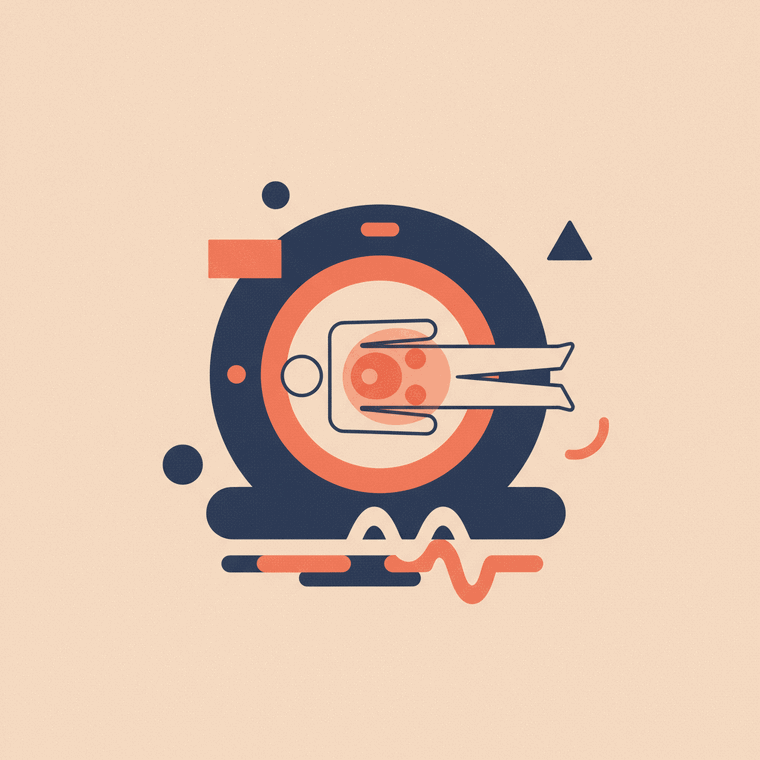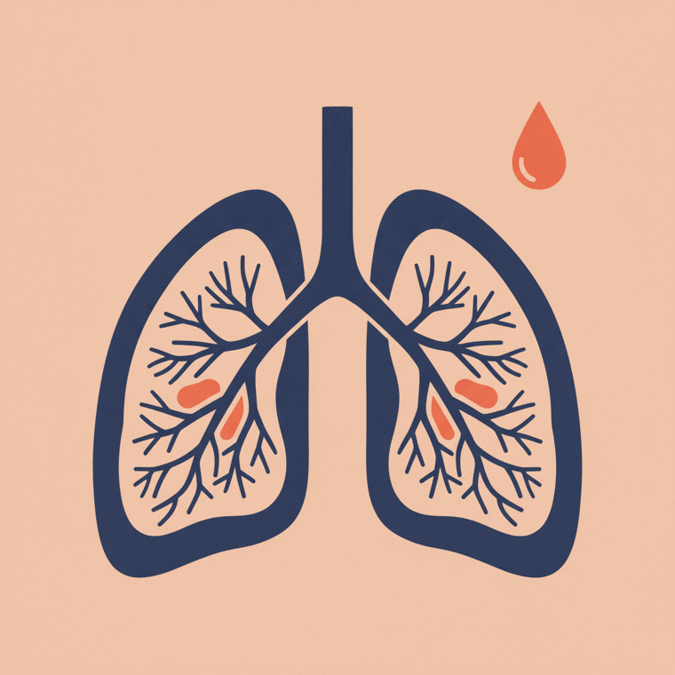
A CT (computed tomography) scan uses X-rays and a computer to build detailed cross-sectional 'slices' of the body — far more detail than a plain X-ray, and very fast. It's a workhorse for the chest, abdomen and pelvis, for trauma and emergencies, and for detecting and staging cancer. A scan is painless and often takes just a few minutes. It does use radiation (more than an X-ray), so it's used when the benefit clearly justifies it — and, unlike MRI, implants are no barrier.
Key takeaways
- CT uses X-rays + a computer to build detailed cross-sectional images in seconds.
- It's excellent for the chest, abdomen, pelvis, trauma, and cancer detection.
- It's fast and, unlike MRI, works fine with implanted devices.
- It uses more radiation than an X-ray, so it's used when clearly justified.
If an X-ray is a flat photograph, a CT scan is a set of thin, detailed slices you can stack up and view from any angle. It’s one of the most useful — and fastest — scans in medicine. Here’s how it works and what it’s for.
What a CT scan is
CT stands for computed tomography (you may also hear “CAT scan”). Like an X-ray, it uses radiation — but instead of one flat image, several X-ray beams and detectors rotate around you, and a computer combines the data into detailed cross-sectional slices.[1] A common way to picture it: like looking at a loaf of bread by cutting it into thin slices, then reassembling them into a 3D view.[1]
The result shows bone, soft tissue and blood vessels all at once, in far more detail than a plain X-ray — especially for soft tissue.[1]
What CT is used for
CT is a genuine all-rounder. Common uses include:[1]
- Trauma and emergencies — it’s fast and excellent at spotting internal injuries and bleeding, which can be life-saving.
- Chest, abdomen and pelvis — detailed cross-sections of the lungs, liver, kidneys, bowel and more, for pain, infection or unexplained symptoms.
- Cancer — detecting tumours, measuring their size and location, and staging cancers of the lung, liver, kidney, ovary and pancreas.
- Blood vessels — assessing a suspected pulmonary embolism (clot in the lung) or an aortic aneurysm (a “CT angiogram”).
- Guiding procedures — steering a needle precisely during a biopsy or drainage.
Its speed is a real advantage: modern scanners image large areas in seconds, which suits emergencies, children and anyone who finds it hard to stay still.[1]
What a scan is like
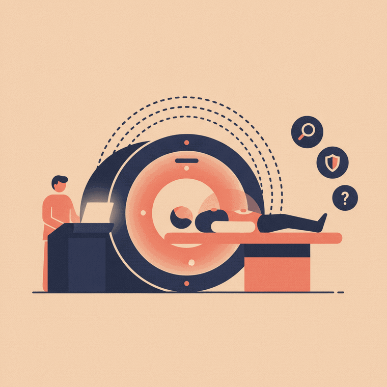
In an Australian radiology practice, a radiographer positions you on a table that slides through the ring-shaped scanner (a short, open “donut”, not a long tunnel).[1] You lie still — sometimes holding your breath briefly — while the table moves through. The whole exam is usually done within about 30 minutes, and the scanning itself is often just a few minutes.[1] It’s painless, and the radiographer watches and talks to you throughout.
Contrast dye
Many CT scans use an iodine-based contrast, usually injected into a vein, to make blood vessels and organs stand out.[2] As it goes in, it’s normal to feel a warm flush and a metallic taste — this passes quickly. Some scans use a contrast drink instead, to outline the bowel. (See our guides on preparing for a CT and CT contrast safety.)
CT vs MRI: which is which
They’re complementary, not rivals:[1]
- CT is faster, better in emergencies, and superb for the lungs, bone detail and acute bleeding — and it works with any implant.
- MRI shows soft tissue (brain, spine, joints) in more detail and uses no radiation, but is slower and has strict metal rules.
Your doctor chooses based on the question, the urgency, and keeping radiation as low as reasonable.
Getting your results
A radiologist interprets the images and sends a report to your referring doctor, who explains what it means. You won’t get results from the radiographer on the day.[1]
Frequently asked questions
Is a CT scan painful?
No — it’s painless. The only minor discomforts are lying still, and (if you have contrast) the pinprick of the injection and a brief warm flush.[1]
How long does a CT scan take?
The scan itself is often just a few minutes; the whole appointment is usually within about 30 minutes, longer if contrast is used.[1]
Can I have a CT scan if I have a pacemaker or metal implant?
Yes. Unlike MRI, an implanted device of any kind won’t stop you having a CT scan — one of CT’s practical advantages.[1]
Does a CT scan use a lot of radiation?
More than a plain X-ray, but the dose varies with the scan, and modern scanners minimise it. It’s used when the benefit outweighs the small risk. (See our guide to CT risks and radiation.)[4]
Sources
- RadiologyInfo.org (RSNA & ACR) — CT of the Body — www.radiologyinfo.org/en/info/bodyct
- RANZCR / InsideRadiology — Computed tomography (CT) — www.insideradiology.com.au/computed-tomography/
- healthdirect — CT scan — www.healthdirect.gov.au/ct-scan
- ARPANSA — Radiation and health / medical imaging doses — www.arpansa.gov.au/understanding-radiation/radiation-sources/more-radiation-sources/medical-imaging