A DXA (bone mineral density) scan is the gold-standard test for measuring bone strength and diagnosing osteoporosis. It's quick, painless and very low-dose — less than a tenth of a chest X-ray, and under a day of natural background radiation. Results are given as a T-score: above −1 is normal, −1 to −2.5 is osteopenia (mild bone loss), and below −2.5 is osteoporosis. The only absolute contraindication is pregnancy.
Key takeaways
- DXA is the gold-standard scan for bone density and osteoporosis.
- It's quick, painless and uses a very low dose of radiation.
- Results are a 'T-score': above −1 normal, −1 to −2.5 osteopenia, below −2.5 osteoporosis.
- Pregnancy is the only absolute contraindication; DXA is safe with a pacemaker.
A bone mineral density (BMD) scan — usually called a DXA scan — is the gold-standard test for measuring how strong your bones are and diagnosing osteoporosis (thinning of the bones that raises fracture risk).[1] It’s a quick, painless, very low-dose X-ray, most often of the hip and lower spine.
Who needs a DXA scan?
A DXA is used for people at increased risk of bone loss and fracture. Common reasons include:[2]
- Being aged 70 or over.
- Having had a low-impact fracture (a break from a fall at standing height) or a vertebral fracture.
- Conditions or medications that can cause bone loss — for example long-term corticosteroids, an overactive thyroid, rheumatoid arthritis, coeliac or Crohn’s disease, or chronic kidney or liver disease.
Some of these reasons attract a Medicare rebate; others may still be worthwhile but aren’t rebatable. Your doctor will advise.[2]
What your T-score means
Results are reported as a T-score, comparing your bone density to a healthy young adult of the same sex (World Health Organization criteria):[1]
| T-score | Meaning |
|---|---|
| Above −1 | Normal |
| −1 to −2.5 | Osteopenia (mild bone loss) |
| Below −2.5 | Osteoporosis |
A second number, the Z-score, compares you with others of your own age, size and sex; an unusually high or low Z-score can prompt further tests.[1] Your doctor interprets the scores alongside your overall health and other risk factors.
What to expect, and is it safe?
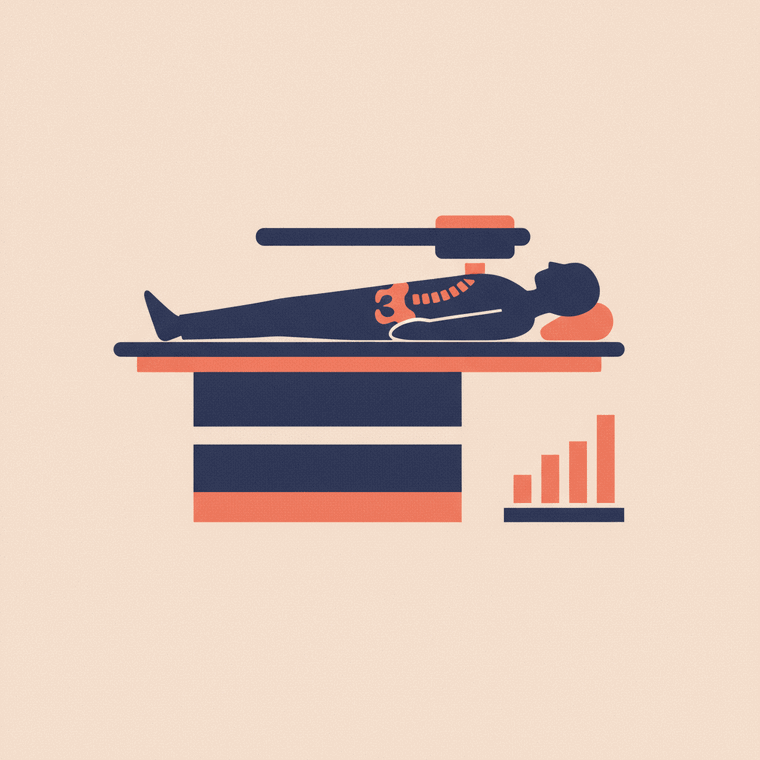
You lie on a table while a scanning arm passes over you — it’s painless, needs no injection, and there’s no aftercare.[1] A DXA uses a very low dose of radiation — less than a tenth of a chest X-ray, and under a day of natural background radiation.[1]
A few practical points:[2]
- Pregnancy is the only absolute contraindication (because of the radiation) — tell staff if you could be pregnant.
- DXA is safe for people with a pacemaker or defibrillator.
- Metal implants like a hip replacement or spinal fusion mean that site can’t be scanned, so another site is used.
- Avoid a DXA for 10–14 days after a scan that used contrast or a nuclear medicine tracer, as these can affect the result.[1]
- Don’t take calcium supplements for 24 hours beforehand.[1]
For monitoring over time, follow-up scans should ideally be on the same scanner, because machines from different makers can’t be directly compared.[1]
Frequently asked questions
Does a DXA scan hurt or involve an injection?
No — it’s painless, needs no injection, and there’s minimal preparation and no aftercare. You simply lie still while the scanner passes over you.[1]
Is a DXA scan safe?
Yes. It uses a very low dose of radiation — less than a day of natural background — and has no apparent adverse effects. The only absolute contraindication is pregnancy.[1]
How often should I have a DXA?
Often every 2 years to see a meaningful change, though people on high-dose steroids may be checked sooner. Your doctor sets the interval based on your result and risk.[1]
Sources
- RadiologyInfo.org (RSNA & ACR) — Bone Density Scan (DEXA/DXA) — www.radiologyinfo.org/en/info/dexa
- RANZCR / InsideRadiology — Bone mineral density scan (DXA) (Hendrich) — www.insideradiology.com.au/bone-mineral-density-scan-hp/