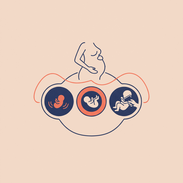
The general approach to imaging stays the same throughout pregnancy — favour radiation-free tests, and weigh benefit against a small risk — but caution is greatest in the first trimester, when the baby is developing most rapidly. Ultrasound is used safely at every stage. MRI is used carefully in the first trimester; radiation-based scans are used at any stage only when genuinely necessary.
Key takeaways
- The core principles are the same all through pregnancy.
- Caution is greatest in the first trimester.
- Ultrasound is used safely at every stage.
- MRI is used carefully in the first trimester; radiation scans only if needed.
The principles of imaging in pregnancy don’t change from week to week — favour radiation-free tests, and weigh benefit against a small risk. But the degree of caution does shift, mainly because of how the baby develops.[3]
First trimester — the most cautious
Early pregnancy is when the baby is developing most rapidly, so caution is greatest:
- Ultrasound is used safely (it’s how an early dating scan is done).[2]
- MRI is used carefully in the first trimester — reserved for when it’s needed and discussed with the radiologist. The risks are considered theoretical and probably small, but aren’t fully established.[2]
- X-ray and CT are avoided unless genuinely necessary, with the lowest dose and the beam focused away from the baby if used.
Second and third trimesters
The same priorities apply, with the first-trimester caution around MRI easing. Ultrasound remains the mainstay (including the 18–20 week anatomy scan). MRI may be used when ultrasound raises a question. Radiation-based scans are still used only when needed, balanced against the benefit.[3]
What doesn’t change
At every stage: tell your team you’re pregnant, expect ultrasound or MRI to be preferred, and know that gadolinium contrast is usually avoided throughout (while iodine contrast isn’t known to harm the baby).[1]
Frequently asked questions
Is imaging more risky early in pregnancy?
Caution is greatest in the first trimester because the baby is developing fastest. Ultrasound is safe throughout; MRI is used carefully early on.[2]
Does the safe approach change later in pregnancy?
The core principles stay the same. The main shift is that the extra first-trimester caution around MRI eases later on.[3]
Sources
- RadiologyInfo.org (RSNA & ACR) — Contrast Materials (pregnancy) — www.radiologyinfo.org/en/info/safety-contrast
- RANZCR / InsideRadiology — Magnetic resonance imaging (MRI) — www.insideradiology.com.au/mri-hp/
- RANZCR / InsideRadiology — Radiation risk of medical imaging — www.insideradiology.com.au/radiation-risk/