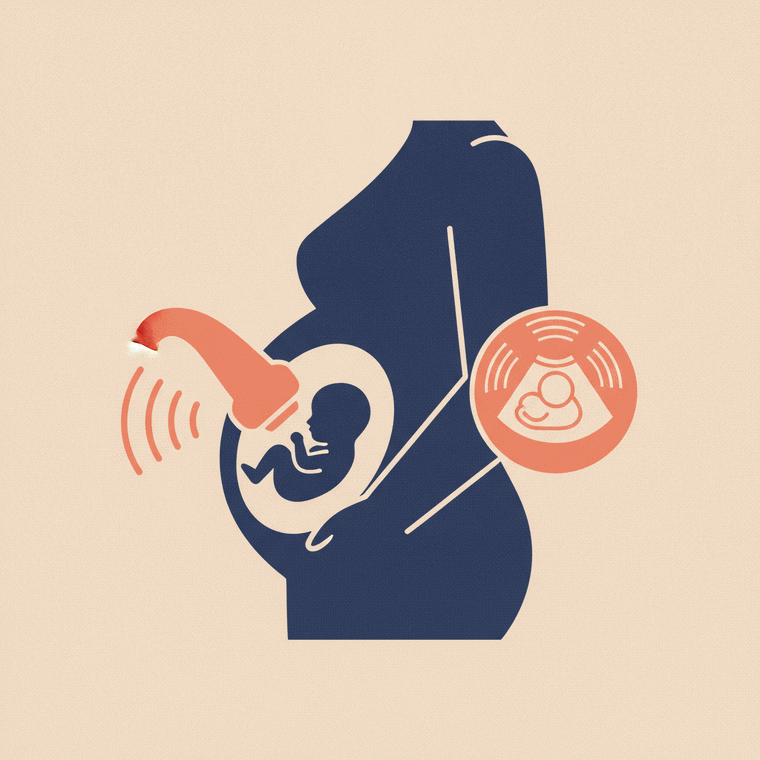
A pregnancy ultrasound uses sound waves — no radiation — to check on your baby, and is the standard, safe way to monitor a pregnancy. Common Australian scans include an early dating scan, an 11–13 week scan, and a mid-pregnancy morphology (anatomy) scan around 18–20 weeks. It's painless; some scans need a full bladder or are done internally early on.
Key takeaways
- Ultrasound is the standard, radiation-free way to monitor pregnancy.
- Common scans include an early dating scan and a mid-pregnancy anatomy scan.
- The 18–20 week morphology scan checks the baby's development in detail.
- It's painless and considered safe; some scans need a full bladder.
Ultrasound is the mainstay of pregnancy imaging. Because it uses sound waves rather than radiation and has no known adverse effects, it can be used to monitor your baby safely throughout the pregnancy.[2]
The common scans
While the exact schedule depends on your care, common pregnancy ultrasounds in Australia include:
- An early dating scan, which confirms the pregnancy and estimates how far along you are.
- An 11–13 week scan (the nuchal translucency), sometimes combined with screening for chromosomal conditions.
- A mid-pregnancy morphology (anatomy) scan, usually around 18–20 weeks, which checks the baby’s development in detail — the brain, heart, spine, organs, limbs and the placenta.
- Additional growth or wellbeing scans later, if needed.
Because ultrasound is real-time, the sonographer can watch the baby move and assess blood flow during the scan.[1]
What to expect
Most pregnancy ultrasounds are done over the abdomen, with gel and a hand-held probe; some require a full bladder to give a clear view.[1] Very early scans are sometimes done internally (transvaginally) for a closer look. It’s painless, with no aftercare, and typically takes 20–45 minutes depending on the scan.
Is it safe?
Yes — ultrasound is considered safe for both mother and baby, which is exactly why it’s the standard imaging used in pregnancy.[2] As a precaution, obstetric ultrasound is done by trained operators for a medical reason, rather than purely for “keepsake” (3D/4D) images.
Frequently asked questions
Is a pregnancy ultrasound safe for my baby?
Yes — it uses sound waves rather than radiation and has no known adverse effects, which is why it’s used routinely throughout pregnancy.[2]
Why do I need a full bladder for some pregnancy scans?
A full bladder lifts the uterus and improves the view, giving a clearer image — mostly relevant in early pregnancy. Later scans usually don’t need one.[1]
What does the 18–20 week scan check?
It’s a detailed look at the baby’s anatomy — brain, heart, spine, organs and limbs — plus the placenta and fluid. It’s one of the most important routine scans in pregnancy.[1]
Sources
- RadiologyInfo.org (RSNA & ACR) — General Ultrasound — www.radiologyinfo.org/en/info/genus
- RANZCR / InsideRadiology — Ultrasound (Coombs) — www.insideradiology.com.au/ultrasound-hp/