A head (or brain) CT uses X-rays to build fast, detailed images of the skull and brain. It's the first-line scan in emergencies — head injuries and suspected stroke — because it's quick and very good at detecting bleeding. The scan takes only a few minutes, is painless, and often needs no contrast. It uses a modest radiation dose (about 2 mSv, roughly a year of natural background). For detailed, non-urgent brain assessment, MRI is usually preferred.
Key takeaways
- Head CT is fast and excellent at detecting bleeding — ideal in emergencies.
- It's the first scan for head injuries and suspected stroke.
- It takes minutes, is painless, and often needs no contrast.
- For detailed non-urgent brain problems, MRI usually shows more.
When someone hits their head or a stroke is suspected, the scan that’s usually done first is a CT of the head — and there’s a good reason for that.
Why CT first for the head
A head CT is fast (just a few minutes) and it’s excellent at detecting bleeding inside or around the brain.[1] In an emergency, that speed and sensitivity to blood can be life-saving — which is why CT, not MRI, is the first-line scan for:[2]
- Head injuries — to check for bleeding or a skull fracture
- Suspected stroke — crucially, to rule out a bleed before any clot-dissolving treatment is given
- Sudden severe headache, confusion or loss of consciousness
- Assessing pressure or fluid changes in the brain
What a head CT is like
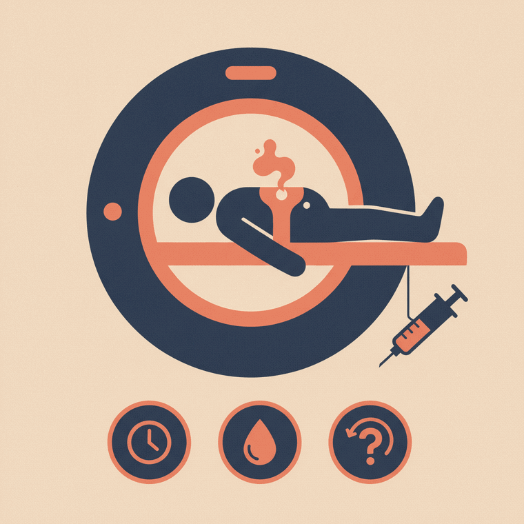
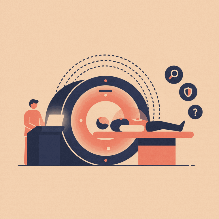
You lie on your back with your head resting in a cradle, and the table moves your head through the ring-shaped scanner.[1] You’ll be asked to keep still; the scan itself takes only a few minutes, and it’s painless.[1] Many head CTs need no contrast at all; if contrast is used (for example, to look at blood vessels or a suspected tumour), it’s injected into a vein and you may feel a brief warm flush.
The radiation
A head CT delivers about 2 mSv — roughly a year of natural background radiation.[3] It’s more than a plain X-ray but a modest CT dose, and in the situations above the benefit clearly outweighs the small risk. In children, low-dose settings are used.
Head CT vs brain MRI
They’re used for different jobs:[1]
- CT is faster and better for emergencies and bleeding — the right first move when time matters.
- MRI shows the brain’s soft tissue in far more detail and uses no radiation, making it better for non-urgent investigation of things like tumours, MS or persistent symptoms.
Often the two are complementary: a CT rules out an emergency, and an MRI follows if more detail is needed.
Frequently asked questions
Why a CT and not an MRI for my head injury?
Because CT is fast and very good at spotting bleeding — exactly what needs ruling out quickly after a head injury. MRI may follow later if more detail is needed.[1]
Does a head CT hurt?
No — it’s painless and takes only a few minutes. If contrast is used you’ll feel the pinprick of the injection and possibly a brief warm flush.[1]
Is the radiation from a head CT dangerous?
The dose (about 2 mSv, roughly a year of background radiation) is modest, and in the situations a head CT is used, the benefit far outweighs the small risk.[3]
Sources
- RadiologyInfo.org (RSNA & ACR) — CT of the Body — www.radiologyinfo.org/en/info/bodyct
- RANZCR / InsideRadiology — Computed tomography (CT) — www.insideradiology.com.au/computed-tomography/
- ARPANSA — Medical imaging radiation doses — www.arpansa.gov.au/understanding-radiation/radiation-sources/more-radiation-sources/medical-imaging