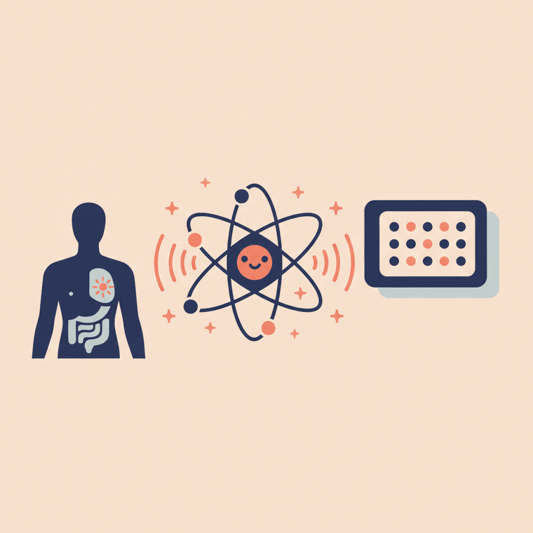
A radioactive tracer (radiopharmaceutical) has two parts: a radioactive label that gives off energy the camera can detect, and a carrier molecule that takes it to the organ being studied. Only tiny amounts are used. The most common general label is Technetium-99m; PET usually uses FDG, a glucose-like tracer that active cells take up. Tracers have a short half-life, so most of the radioactivity is gone within hours to about a day.
Key takeaways
- A tracer has a radioactive label plus a carrier molecule that targets an organ.
- The carrier decides where the tracer goes (bone, thyroid, tumour…).
- Technetium-99m is the common general label; PET uses FDG (glucose-like).
- Tracers have a short half-life — most is gone within hours to a day.
The radioactive tracer — properly called a radiopharmaceutical — is what makes a nuclear medicine scan work. Understanding it takes the mystery (and much of the worry) out of these scans.
The two parts of a tracer
A tracer has two jobs rolled into one molecule:[1]
- A radioactive label — an unstable atom that gives off energy as it decays. This is what the gamma camera or PET scanner detects.
- A carrier molecule — the part that decides where the tracer goes in the body. A bone-seeking carrier heads to the skeleton; a glucose-like carrier goes to metabolically active cells.
So it’s the carrier that targets the organ, and the label that lets the camera see it. Only a tiny amount is used — far too little to have any drug-like effect.[1]
The common tracers
- Technetium-99m is the workhorse of general nuclear medicine, attached to different carriers for different scans (bone, kidney, thyroid, heart).[1]
- FDG (F-18 fluorodeoxyglucose) is the most common PET tracer — a compound similar to glucose (sugar). Because very active cells (including many cancers) use more glucose, they take up more FDG and “light up”.[1]
- Others include radioactive iodine (for the thyroid) and various specialised tracers.
Why it leaves the body quickly
Each radioactive label has a half-life — the time for half of it to decay away. For the labels used in nuclear medicine, this is measured in hours, so after a day or so there’s very little radioactivity left.[1] The tracer is cleared from your body mainly in your urine (and sometimes stool or breath), which is why you’re encouraged to drink plenty of fluids afterwards.
Frequently asked questions
How does the tracer know where to go in my body?
The carrier molecule is chosen to travel to a specific target — a bone-seeking carrier goes to the bones, a glucose-like one to active cells, and so on. The radioactive label just comes along for the ride so the camera can see it.[1]
How long does the tracer stay radioactive?
Not long — its half-life is measured in hours, so most of the radioactivity is gone within about a day, and it’s cleared mainly in your urine.[1]
Is the tracer a drug I could react to?
The amount is tiny — far below a drug dose — and reactions are extremely rare. Still, always mention any allergies or past reactions to the nuclear medicine team.[1]
Sources
- RadiologyInfo.org (RSNA & ACR) — General Nuclear Medicine — www.radiologyinfo.org/en/info/gennuclear
- RANZCR / InsideRadiology — Nuclear medicine (Cain) — www.insideradiology.com.au/nuclear-medicine/