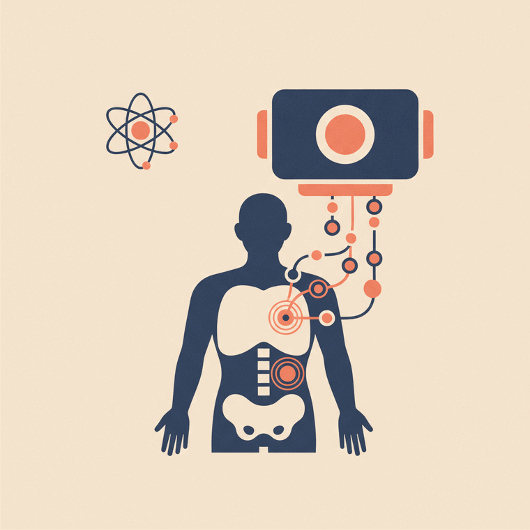
A bone scan highlights areas of increased bone activity — 'hot spots' where the bone is busy repairing or reacting. A hot spot can mean many things: a healing fracture, arthritis, infection, or cancer spread — so it's a clue, not a diagnosis. A specialist reads the whole pattern, and often recommends a follow-up X-ray, CT or MRI of a hot spot to pin down the cause, before reporting to your doctor.
Key takeaways
- A bone scan highlights areas of increased (or decreased) bone activity.
- A hot spot can be a fracture, arthritis, infection or cancer — a clue, not a diagnosis.
- The pattern and location of hot spots guide what they likely mean.
- Follow-up imaging of a hot spot (X-ray, CT, MRI) is common.
If you’ve had a bone scan, the report may mention “hot spots” or “increased uptake.” That can sound alarming — so here’s what it actually means.
What a hot spot is
A bone scan shows how active different parts of your skeleton are. Areas of increased activity — where the bone is busy repairing or reacting to something — show up as “hot spots.”[1] (“Cold spots,” areas of reduced activity, can also be meaningful.)
A hot spot is a clue, not a diagnosis
The key thing to understand: increased bone activity has many possible causes, so a hot spot is a clue, not a verdict. It can come from:[1]
- A healing fracture (including small stress fractures)
- Arthritis or wear-and-tear
- Infection in the bone
- Cancer that has spread to bone
The pattern and location of the hot spots help the specialist work out the likely cause — for example, the symmetrical pattern of arthritis looks different from the scattered pattern that cancer spread can produce.[2]
What usually happens next
Because a hot spot has several possible causes, the specialist often recommends a closer look at the area with an X-ray, CT or MRI to pin down exactly what’s going on.[1] The radiologist or nuclear medicine specialist who reads the scan writes a report for the doctor who referred you, who explains what it means and the next steps.[1]
Frequently asked questions
Does a hot spot on my bone scan mean cancer?
Not necessarily. Fractures, arthritis and infection all cause hot spots too. The pattern guides interpretation, and a hot spot is often checked with a more detailed scan of that area before any conclusion.[1]
Why do I need another scan after a bone scan?
Because a bone scan shows where activity is, but not always why. An X-ray, CT or MRI of a hot spot gives the detail needed to explain it.[1]
Who explains the results to me?
The scan is interpreted by a specialist and reported to your referring doctor, who discusses what it means for you alongside your symptoms.[1]
Sources
- RadiologyInfo.org (RSNA & ACR) — General Nuclear Medicine — www.radiologyinfo.org/en/info/gennuclear
- RANZCR / InsideRadiology — Nuclear medicine (Cain) — www.insideradiology.com.au/nuclear-medicine/